Motivation and Objective
Non-contrast chest computed tomography (CT) has been used for monitoring of the healthy status, mainly in China, of COVID-19 patients with a significant success. However, this approach cannot be used massively mainly for high risk, cost and limited availability being this tool not extensively available in some countries. In addition, when used for evaluating COVID-19 patients, difficult and time-consuming procedures of decontamination have to be set after each scan causing both increasing cost for patient and reducing operation time of the CT.
Alternatively, chest X-ray (CXR), although less sensitive than CT-scan, can provide important information about the evolution of pulmonary involvement during the disease to verify the response of a patient to treatments. According to recent guidelines of North American Radiology Scientific Expert Panel, portable CXR has to be considered as the main imaging approach in evaluating COVID-19 patients [1] which not only reduces radiations for patients, but also avoids their transportation and provides the possibility to monitor patients in the intensive care units (ICUs) which are more than 5% of the total known cases of COVID-19.
Those images are often of low quality for the environmental difficulties and for non-collaborative and severely ill patients, in most cases causing many different artifacts originating inhomogeneities in luminance distribution of radiograms.
EX-COVID will address this problem by developing a tool, named PACE (Pipeline for Advanced Contrast Enhancement), which will support radiologists in evaluating low quality CXRs for monitoring the healthy status of COVID-19 patients.
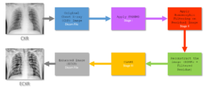
The start point of EX-COVID is the tool Pipeline for Advanced Contrast Enhancement (PACE) which block diagram is shows in the figure. As step one, the CXR is processed with the Fast and Adaptive Bidimensional Empirical Mode Decomposition (FABEMD) which decomposes the input image into multiple hierarchical components named bidimensional intrinsic mode functions (BIMFs) and a bidimensional residual image (BR). The residual image is filtered with a Homomorphic Filter (HMF) with a High-Frequency Emphasis Filter (HEF) as kernel function. The CXR is then reconstructed combining the BIMFs with the filtered residual image.

Zoom